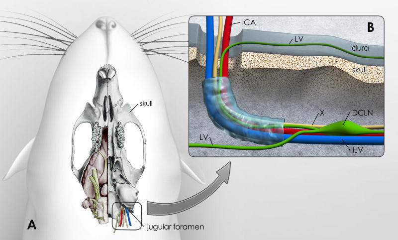
Fig. 5. Vessels, cranial nerves and lymph vessels at the site of the jugular foramen.
Fig. 5 is an illustration of how lymphatic vessels in the dura might drain to the deep cervical lymph nodes (DCLN). A: The ventral aspect of the rat skull is shown; the right side shows the brain in situ (modified from: [85]). The jugular foramen is highlighted and shows the exit of the vagal nerve (X), internal jugular vein (IJV) and internal carotid artery (ICA). B: High magnification of the area of the jugular foramen with the proposed exiting vessels and vagal nerve. Lymphatic vessels (LV) associated with dura lining the ventral surface of the skull might exit at this site into the DCLNs. This drawing is a proposed illustration of the connection between dural LVs and DCLNs.

