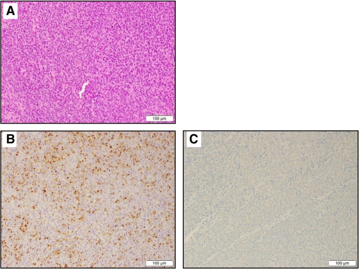
Fig. 3.
a Histopathological findings at the section, which is located on the yellow line in Fig. 2c (hematoxylin and eosin staining). The tumors were located at the submucosa and exhibited hyperplasia-like epithelioid cells but no melanocytes. Histopathological findings (immunohistochemical staining). b The tumors were diffusely positive for HMB45. c The tumors were partially positive for Melan-A

