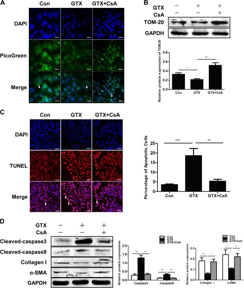
Fig. 2. Inhibition of mitophagy suppresses apoptosis in HSCs.
a Mitochondrial DNA (mtDNA) measured by PicoGreen staining in LX2 cells; Scale bar, 10 μm. b Representative western blots of TOM20 with GAPDH serving as the internal reference. Bar graph represents the mean ± SEM. *P < 0.05 and **P < 0.01 vs. the indicated groups. c TUNEL staining of LX2 cells from the indicated groups; scale bar, 25 μm. Bar graph represents the mean ± SEM. **P < 0.01 and ***P < 0.001 vs. the indicated groups. d Representative western blots of cleaved caspase3, cleaved caspase9, collagen I and α-SMA. Bar graph represents the mean ± SEM of three different experiments. *P < 0.05 and **P < 0.01 vs. the indicated groups

