Fig. 2.
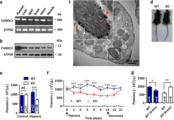
FUNDC2 knockout mice display hypoxia-induced thrombocytopenia. a The RNA of indicated mouse tissues was extracted and the product of reverse transcription-PCR was shown. b Expression profiling of mouse tissues for FUNDC2. WAT white adipose tissue. Same mitochondrial content in each sample. c Immunogold electron micrograph of FUNDC2 in platelets. Insert: enlarged view of immunogold particles conjugated with antibodies against Nix (mitochondrial outer membrane protein, 10 nm particles, white arrow) or FUNDC2 (20 nm particles, red arrowheads). d FUNDC2 knockout (KO) mouse and the wild-type (WT) littermate. e Platelet counts were determined from mice treated with hypoxia for 5 days. f Mice were exposed to hypoxia and then put back in normoxia for indicated days and platelet counts were determined (n = 5–10 mice). g Transplantation (trans) of bone marrow. Mice were treated with hypoxia for 5 days (n = 6 mice)

