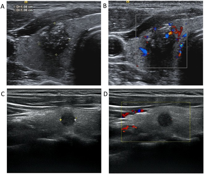
Figure 2.
Ultrasonographic images of a PTC patient with (A,B) and without (C,D) cervical lymph node metastasis. (A) A nodule with certain malignant signs, such as hypoechogenicity, irregular shape, ill-defined margin, multiple microcalcification and capsule invasion; (B) CDFI showed rich blood flow in and around the nodule (A). (C) A hypoechoic nodule in the middle of the right thyroid, smaller than that in (A), with regular shape, defined margin, and no signs of capsule invasion; (D) CDFI showed lack of the blood flow signals, compared with that in (B). CDFI: Color Doppler Flow Imaging.

