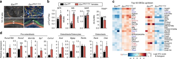
Fig. 3.
Increased bone formation in Esr1Nkx2-1Cre females. a Representative images of labeled mineralized surface of distal femur with calcein (green) and demeclocycline (orange) over a period of 1 week in a 12 week old female. Scale bars = 50 μm. b Dynamic histomorphometric results for Esr1fl/fl (n = 4, black bars) and Esr1Nkx2-1Cre (n = 4, red bars) 12–14 week females showing bone formation rate (BFR), mineralized surface (MS), and mineralized apposition rate (MAR). Number of active osteoclasts normalized to bone surface quantified by TRAP-positive staining determined in distal femurs from 5- to 7-week-old Esr1fl/fl (n = 5) and Esr1Nkx2-1Cre (n = 6) females. c Heat map of top 50 differentially expressed genes (DEGs) up and down in 4.5-week-old Esr1fl/fl and Esr1Nkx2-1Cre bone marrow Esr1fl/fl (n = 4) and Esr1Nkx2-1Cre (n = 4) females. BMP regulated genes (red) and IFN regulated genes (blue). d Quantification of indicated transcripts marking pre-osteoblasts, osteocytes, and osteoclasts in 4.5–7-week female control (n = 10) and mutant (n = 7) flushed bone marrow (BM), or in female control (n = 13) and mutant (n = 8) femur bone chips. Error bars are ±SEM. Unpaired Student’s t test (b, d). *p < 0.05; ****p < 0.0001. NS = p > 0.05

