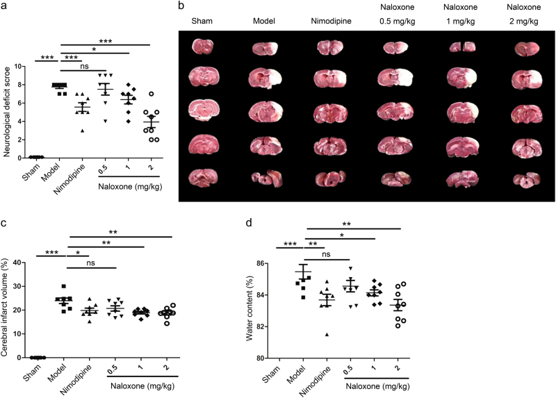
Fig. 1.
Effect of naloxone on ischemia-induced motor deficits and brain injury in rats. a Motor deficits measured 24 h after ischemic insult. Nimodipine was used as a positive control. b Representative photographs of coronal sections with 2% 2,3,5-triphenyltetrazolium chloride staining at 24 h after ischemia in the sham, model, nimodipine, and naloxone rat groups. The white regions are defined as infarct regions. c Quantitative analysis of the total lesion volume in the rat brains. d Water content measured 24 h after ischemic insult. The columns and bars represent the mean ± SEM; n = 8; *P < 0.05; **P < 0.01; ***P < 0.001. ns not significant

