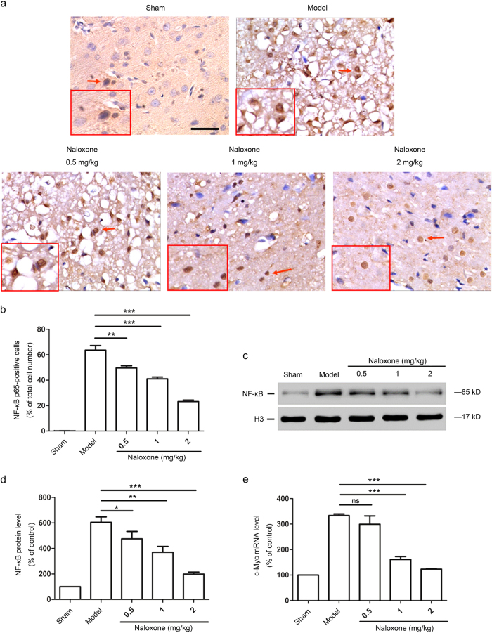
Fig. 3.
Naloxone blocked the nuclear translocation and expression of NF-κB p65 in the ischemic penumbra 12 h after ischemia. a Representative photomicrographs showing the immunohistochemistry of NF-κB p65 in the ischemic penumbra after pMCAO. Cells with positive NF-κB p65 immunoreactivity in the nuclei (arrowheads) were counted as nuclear NF-κB p65-positive cells. The insets show high-magnification views. Scale bar = 50 μm (b) Quantitative analysis of NF-κB p65-positive cells in the ischemic penumbra. The data are expressed as the percentage of the total number of cells. Results are mean ± SEM (n = 8 for each group); **P < 0.01; ***P < 0.001. c Western blot analysis of NF-κB p65 in the nuclear lysates from the ischemic penumbra after pMCAO. The expression of H3 was used as an internal protein loading control. d Quantitative analysis of the changes in NF-κB p65 protein levels in the nuclei from the ischemic penumbra. The data are normalized to the loading control H3 and are expressed as a percentage of the levels in sham-operated animals. The results are the mean ± SEM; n = 3; *P < 0.05; **P < 0.01; ***P < 0.001. e Real-time PCR analysis of c-Myc mRNA expression levels in the ischemic penumbra. The data are normalized to the loading control GAPDH and are expressed as a percentage of the levels in sham-operated animals (mean ± SEM, n = 3); ***P < 0.001. ns not significant

