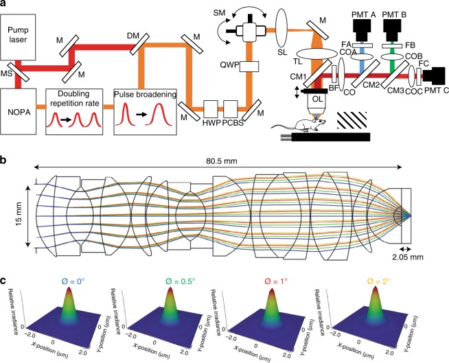
Fig. 1.
Schematic of upright three-photon microscope and layout of the modeled objective lens. a Femtosecond laser pulses from the pump laser (1045 nm) were used to pump the noncollinear optical parametric amplifier (NOPA) to obtain 1300 nm excitation wavelength. The pulse width was elongated using a two-prism pair system and the repetition rate was doubled to 800 kHz by adding a delay line. Power control of laser pulses was performed using a combination of a half-wave plate (HWP) and polarizing cube beam splitter (PCBS). A quarter-wave plate (QWP) was used to control the polarization state of the laser pulses to maximize third harmonic generation (THG) signal on the mouse brain. Laser beams were scanned by a pair of galvanometric scanning mirrors (SM), which were then imaged on the back aperture of a 1.05 NA, 25× objective lens (OL) by a pair of a scan lenses (SL) and a tube lens (TL). Mice were placed on a two-axis motorized stage. The objective was placed on a one-axis motorized stage. Emitted light was collected by a dichroic mirror (CM1, FF705-Di01,Semrock; CM2, Di02-R488, Semrock; CM3, FF555-Di03, Semrock), collection optics (CO), laser blocking filters (BF, FF01-670/SP), and nonlinear imaging filters (FA, FF01-433/24-25, Semrock; FB, FF03-525/50, Semrock; FC, FF01-630/92) and corresponding collection optics (COA, COB, and COC) for each photomultiplier tube (PMT A, PMT B, and PMT C). To make laser ablation marks at different depths of cortex, the output of the pump laser (1045 nm) was sent to the microscope using a mechanical shutter (MS) and a long pass dichroic mirror (DM, Di02-R1064, Semrock). b The optical layout of the objective modeled in Zemax at 1300 nm. The clear aperture of the objective was 15 mm and the working distance was 2.05 mm while using seawater as a sample. c Huygens point spread function results for 4 different scanning angles (0°, 0.5°, 1°, and 2°) on the objective. Numerical apertures in all cases were 1.02 and Strehl ratio values decreased from 0.997 to 0.979 while increasing the scanning angle

