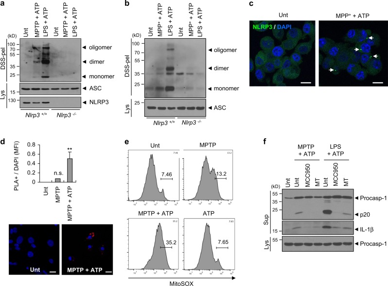
Fig. 4.
MPTP or MPP+ treatment promotes the assembly of the NLRP3 inflammasome. a Immunoblots of disuccinimidyl suberate (DSS)-crosslinked pellets (DSS-pel) or cellular lysates (Lys) from microglia treated with MPTP (40 μM, 16 h) or LPS (0.25 μg ml−1, 3 h), followed by the treatment of ATP (2 mM, 45 min). b Immunoblots of DSS-crosslinked pellets (DSS-pel) or cellular lysates (Lys) from BMDMs treated with MPP+ (100 μM, 6 h) or LPS (0.25 μg ml−1, 3 h), followed by the treatment of ATP (2 mM, 45 min). c Confocal images of NLRP3-GFP-expressing BMDMs untreated or treated with MPP+ (50 μM, 10 h) in the presence of ATP (2 mM, 30 min). Arrows indicate speck-like aggregates of NLRP3 protein (green). DAPI represents the nuclear signal (blue). Scale bars, 10 μm. d Proximity ligation assay of NLRP3 and ASC in mixed glial cells treated with MPTP (40 μM, 16 h), followed by the treatment of ATP (2 mM, 30 min). Proximity ligation (PL) signals (red) represent the molecular association of NLRP3 and ASC. Data are shown as a representative image from four or five independent samples (lower panel). Scale bars, 10 μm. The relative intensity of PL signals (per DAPI signals) was determined and is displayed in the upper panel (n = 4, 5). Data were expressed as the mean ± SEM. Asterisk indicates significant differences (**P < 0.01, n.s. not significant). e Flow cytometric analysis of mixed glial cells treated with MPTP (40 μM, 18 h) and ATP (2.5 mM, final 30 min) after staining with MitoSOX. f Immunoblots from mixed glial cells treated with MPTP (40 μM, 16 h) or LPS (0.25 μg/ml, 3 h) in the presence of MCC950 (50 nM) or Mito-TEMPO (MT, 200 μM), followed by ATP (2.5 mM, 30 min) treatment. a, b, f Culture supernatants (Sup), cellular lysates (Lys), or DSS-crosslinked pellets (DSS-pel) were analyzed by immunoblot

