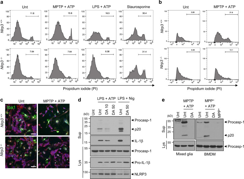
Fig. 6.
Microglial inflammasome activation mediates the death of dopaminergic neurons in a NLRP3-dependent manner. a Flow cytometric analysis of co-cultured microglia and SH-SY5Y cells treated with MPTP (40 μM, 16 h) or LPS (0.25 μg ml−1, 3 h), followed by the treatment with ATP (2.5 mM, 15 min), replenished with fresh medium and incubated for additional 24 h, or treated with staurosporine (1 μg ml−1, 16 h) after staining with anti-CD45 antibody and PI. The PI histograms of CD45-negative SH-SY5Y cells were displayed. b Flow cytometric analysis of co-cultured microglia and MN9D cells treated with MPTP (40 μM, 16 h), followed by the treatment with ATP (2.5 mM, 1 h) after staining with anti-CD45 antibody and PI. The PI histograms of CD45-negative MN9D cells were displayed. c Representative immunofluorescence images of co-cultured microglia and SH-SY5Y cells treated with MPTP (40 μM, 16 h), followed by treatment with ATP (2.5 mM, 15 min) after staining with anti-Iba1 (green) and anti-TH (red) antibodies. DAPI represents the nuclear signal (blue). Scale bars, 50 μm. d Immunoblots from mixed glial cells treated with LPS (0.25 μg ml−1, 3 h) in the presence of dopamine (10 or 50 μg ml−1, 30 min pretreatment before LPS), followed by ATP (2.5 mM, 30 min) or nigericin (Nig, 5 μM, 45 min) treatment. e Immunoblots from mixed glial cells (left) or BMDMs (right) treated with MPTP (40 μM, 16 h, left) or MPP+ (100 μM, 6 h, right) in the presence of dopamine (50 μg ml−1, 30 min pretreatment), followed by ATP (2 mM, 40 min) treatment. Culture supernatants (Sup) or cellular lysates (Lys) were immunoblotted with the indicated antibodies

