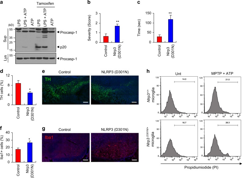
Fig. 8.
Conditional expression of NLRP3 active mutant in microglia exacerbates motor dysfunctions and dopaminergic neuronal loss in MPTP-treated mice. a Immunoblots from Cx3Cr1Cre-ER/+Nlrp3D301NneoR/+ mouse mixed glial cells treated with tamoxifen (1 μM, 24 h), replenished with the medium containing LPS (0.25 μg ml−1, 3 h), followed by ATP (2 mM, 40 min). b–g Control (Cx3Cr1Cre-ER/Cre-ERNlrp3+/+) or NLRP3 mutant mice (Cx3Cr1Cre-ER/+Nlrp3D301NneoR/+) were treated with tamoxifen for 5 days, followed by the administration of MPTP for additional 3 days. b The severity of hindlimb clasping in MPTP-treated control or Nlrp3 (D301N)-expressing mice. (control, n = 8; Nlrp3 (D301N), n = 7). c The latent time to hold the bar in the catalepsy test in MPTP-treated control or Nlrp3 (D301N)-expressing mice (control, n = 8; Nlrp3 (D301N), n = 7). d Quantification of relative TH-positive cells per DAPI in the confocal images as shown in (e) (control, n = 5; Nlrp3 (D301N), n = 3). f Quantification of relative Iba1-positive cells per DAPI in the confocal images as shown in (g) (control, n = 5; Nlrp3 (D301N), n = 3). e, g Representative immunofluorescence image of the fixed brain sections containing the SN of MPTP-treated control or Nlrp3 (D301N)-expressing mice following tamoxifen treatment after staining with anti-TH antibody (green, e) or anti-Iba1 antibody (red, g). DAPI represents the nuclear signal (blue). Scale bars, 200 μm. h Cx3Cr1Cre-ER/Cre-ERNlrp3+/+ or Cx3Cr1Cre-ER/+Nlrp3D301NneoR/+ mice microglia was treated with tamoxifen (1 μM, 24 h), and co-cultured with SH-SY5Y cells. Flow cytometric analysis of co-cultured Nlrp3+/+ or Nlrp3D301N/+ microglia and SH-SY5Y cells treated with MPTP (40 μM, 16 h), followed by the treatment with ATP (2.5 mM, 15 min), replenished with fresh medium and incubated for additional 24 h, after staining with anti-CD45 antibody and PI. The PI histograms of CD45-negative SH-SY5Y cells were displayed. Data were expressed as the mean ± SEM. Asterisks indicate significant differences (*P < 0.05, **P < 0.01)

