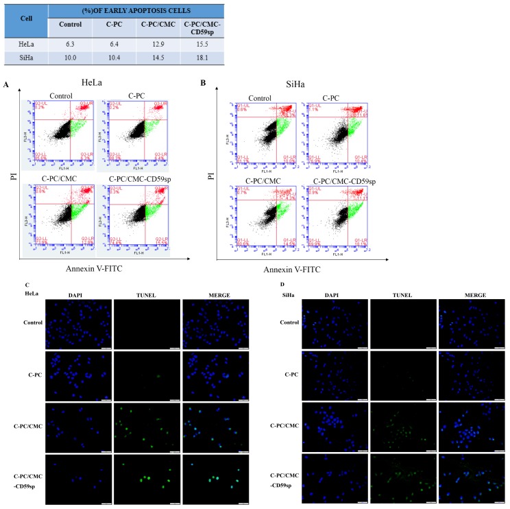
Figure 3.
Effect of nanoparticles on cell apoptosis in HeLa and SiHa cells. (A)Analysis of HeLa cells apoptosis by flow cytometry using Annexin V-FITC and PI after treatment with C-PC, C-PC/CMC NPs and C-PC/CMC-CD59sp NPs for 24h. (B)Analysis of SiHa cells apoptosis by flow cytometry using Annexin V-FITC and PI. Quantitative representation of early apoptotic cells after treatment for 24h. (C) Analysis of HeLa cells apoptosis by TUNEL assay after treatment with C-PC, C-PC/CMC NPs and C-PC/CMC-CD59sp NPs for 24h. (D) Analysis of HeLa cells apoptosis by TUNEL assay. Green fluorescence represents TUNEL-positive cells after treatment.

