Fig. 4.
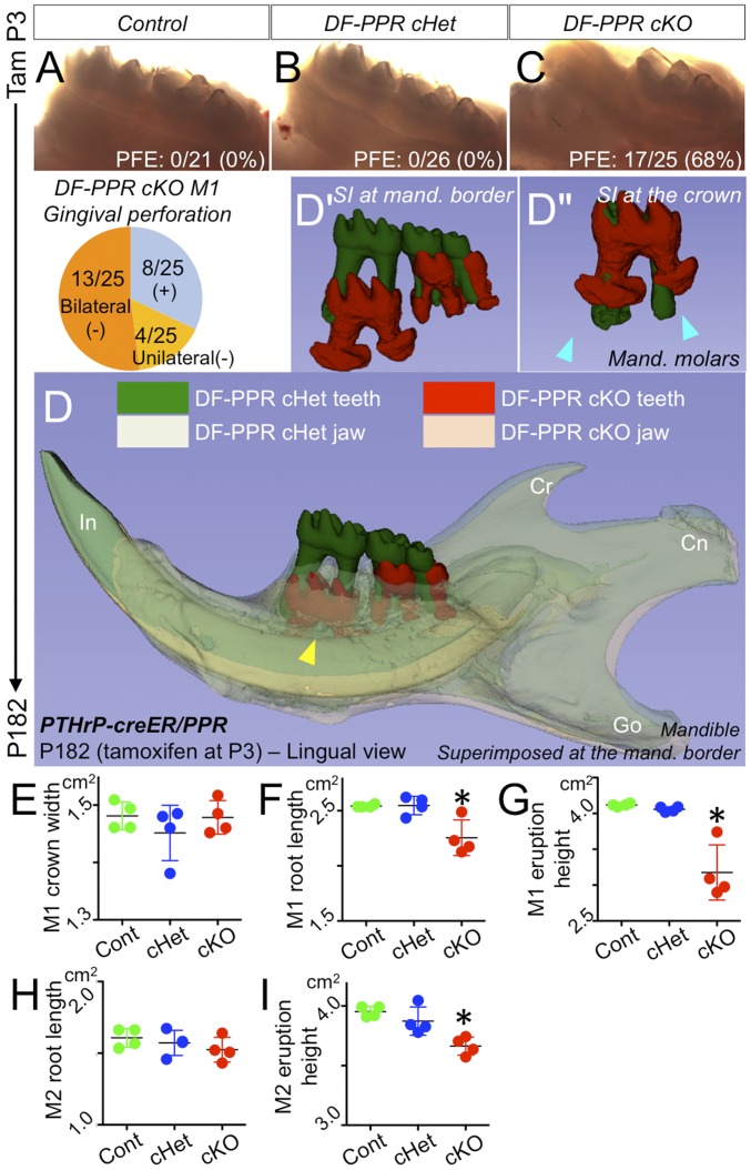
PPR deletion in PTHrP-creER+ DF cells recapitulates human primary failure of eruption conditions. (A–C) PFE phenotypes at 6 mo. (Pie chart) Incidence of failure of eruption (lack of gingival perforation). (D) 3D micro CT surface model overlays: superimposition of registered DF-PPR cHet and DF-PPR cKO mandibles. The yellow arrowhead indicates the first molar associated with pronounced PFE phenotypes and tooth root anomalies. Cn, condyle; Cr, coronoid process; Go, gonial angle; In, incisor. (D′ and D″) Composite 3D surface model overlay of the mandibular first molars. Blue arrowheads indicate truncation (short roots) associated with dilacerations (curved roots). (F–I) Quantitative 3D micro CT analysis. *P < 0.05, one-way ANOVA followed by the Mann–Whitney U test. All data are mean ± SD.

