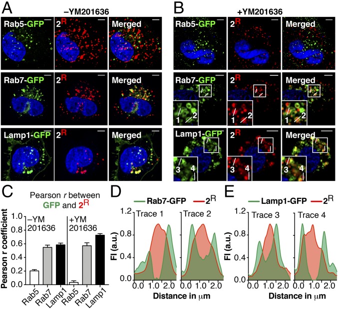
Fig. 4.
CPMP 2 localizes to the lumen of LEs and LYs. Saos-2 cells were transduced with Rab5-, Rab7-, or Lamp1-GFP for 18 h using CellLight Reagents (BacMam 2.0). Cells were washed, incubated with CPMP 2R (300 nM), stained with Hoechst 33342, lifted with TrypLE, and replated into microscopy slides. Cells were then incubated in media ±YM201636 for 1 h. (A and B) Representative live-cell confocal fluorescence microscopy images of Saos-2 cells in media without (A) or with (B) YM201636. (Scale bars: 5 μm.) (C) Pearson correlation coefficients characterizing colocalization of GFP markers and 2R in the presence and absence of YM201636. (D and E) Fluorescence intensity line profiles of endosomes 1–4 (displayed in B) showing the relative location of emission due to 2R (red) and either (D) Rab7-GFP or (E) Lamp1-GFP (green).

