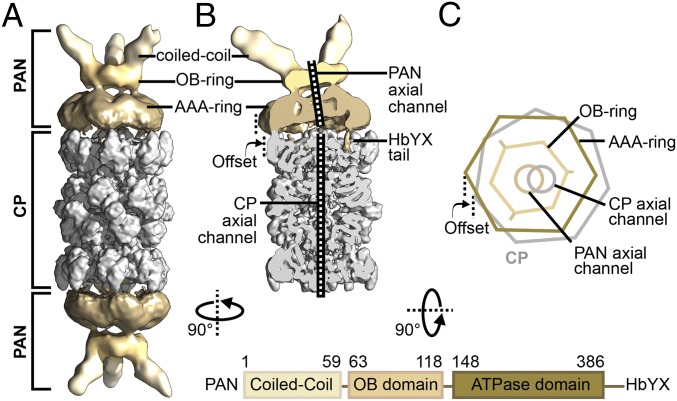
Fig. 1.
Architecture of the PAN-proteasome. (A) Cryo-EM reconstruction of double-capped PAN-proteasomes, with applied C2 symmetry. (B) Cutaway view of PSC particle reconstruction. The density maps in both A and B are filtered according to local resolution, with the CP shown in gray and PAN shown in sand. (C) Simplified illustration of a PAN-proteasome top view. The sevenfold symmetric CP is shown in gray, while the different domains of homohexameric PAN are shown in shades of sand. Mismatch of symmetry between PAN and CP leads to an off-axis location of PAN on top of the CP and the axial pores of PAN and CP are clearly misaligned. The domain architecture of PAN is depicted below.

