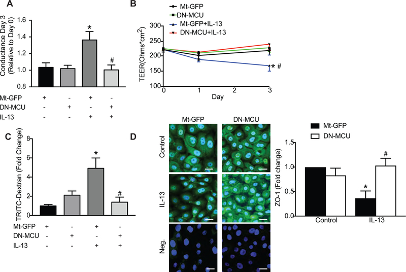
Figure 4. MCU inhibition in HAEC decreases IL-13-mediated conductance and epithelial monolayer permeability.

(A, B) HAEC in Transwell inserts were grown to confluence. Fresh medium was added every 2 days until cells excluded basolateral media. Cells were then infected with Mt-GFP or DN-MCU (10 MOI) for 48 hrs and TEER (ohm × cm2) was monitored after IL-13 treatment (72 hr, 10 ng/ml). Conductance was calculated based on TEER values. (C) Epithelial permeability was assessed by the amount of TRITC-dextran (1 mg/ml) detected in the media of the basolateral compartment of the Transwell and represented as fold-change over GFP-control cells. (D) Representative immunofluorescent images for ZO-1 (green) in HAEC expressing Mt-GFP or DN-MCU after 72 hr treatment with IL-13 or control (63x). Nuclei TO-PRO (blue), negative staining control without primary antibody for Mt-GFP or DN-MCU. Scale bar 100μm. All data are means ± SEM for 3 independent experiments (n = 3 samples/treatment group). Analysis used one-way ANOVA with Tukey post hoc test. * p < 0.05 vs. control; # p < 0.05 vs. Mt-GFP/IL-13.
