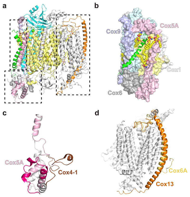
Fig. 2. Structure of S. cerevisiae CIV.
a, Atomic model of CIV showing all 12 subunits. Cox1 is shown in yellow, Cox2 in cyan, Cox5A in pink, Cox26 in green, Cox9 in purple and Cox13 in orange. b, Interactions of Cox26 with Cox1, Cox2, Cox6, Cox9 (coloured as in a) and lipids (spacefill representation). c, Alignment of Cox5A (pink) with its bovine homologue (COX4-1 – grey and brown, PDB 1V54). The first ~50 amino acids are highlighted by a darker shade. d, Differences in length and shape of the transmembrane helix of Cox13 with its bovine homologue (COX6A, PDB 1V54).

