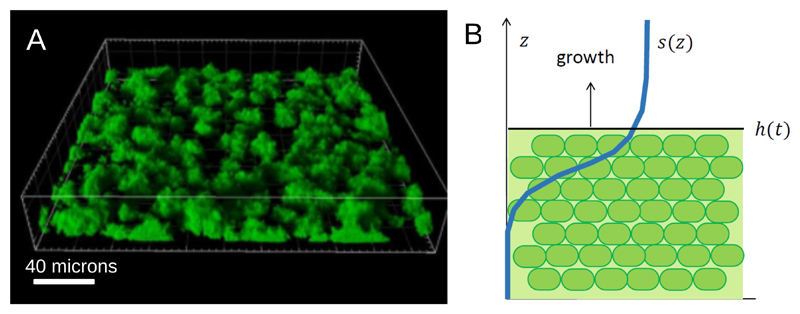
Figure 10.
(A) Confocal laser scanning microscope image of a biofilm formed by P. aeruginosa PAO1 grown for 24 hours in a flow cell. Image reproduced from Ref. [173]. (B) Illustration of a simple model of a growing biofilm with a flat boundary. The position of the boundary is given by z = h(t) and the nutrient profile is s(z).

