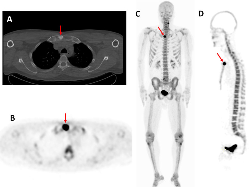
Fig. 5.
A 52 year old man diagnosed with Gleason 4+3 prostate cancer 7 years after radical prostatectomy and salvage radiation with a recent PSA of 2.5 ng/ml. The axial CT image (a) demonstrates an osteoblastic lesion in the sternum (arrow), and the axial (b), coronal (c) and sagittal (d) maximum intensity projection PET images (obtained at 1 h after injection of 18F-NaF) show increased tracer uptake in the sternal osteoblastic lesion (arrows), which is consistent with metastatic disease. Note the uptake in the lateral cervical spine on the left (c), which is degenerative based on its location, indicating the relative lack of specificity of 18F-NaF PET imaging

