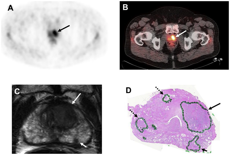
Fig 3.
A 59-year-old man with a prostate-specific antigen (PSA) of 8.6 ng/mL. 11C-acetate positron emission tomography (PET) shows significant tracer uptake in the left-sided lesion on (A) PET and (B) PET/computed tomography images (arrows). (C) Axial T2-weighted magnetic resonance imaging (MRI) shows two lesions (large arrow: left anterior transitional zone; small arrow: left peripheral zone) in the left apical portion of the prostate. (D) Histopathology confirmed the presence of a Gleason 4 + 4 tumour within the left-sided large transitional zone lesion (arrow) and three smaller Gleason 3 + 3 lesions (dashed arrow lesions were missed by both 11C-acetate PET and MRI, whereas the solid arrow lesion was detected by MRI only).

