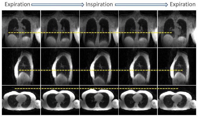
Figure 6.
Motion-resolved lung images during deep breathing (XD-UTE-PF-DB) in a representative subject. Despite reduced image quality and lower spatial resolution compared to normal breathing, images obtained during deep breathing provide dynamic functional information that can be used to obtain pulmonary functional parameters. The varying position of the diaphragm with respect to the yellow dashed lines indicates that respiratory motion can be resolved using XD-UTE reconstruction.

