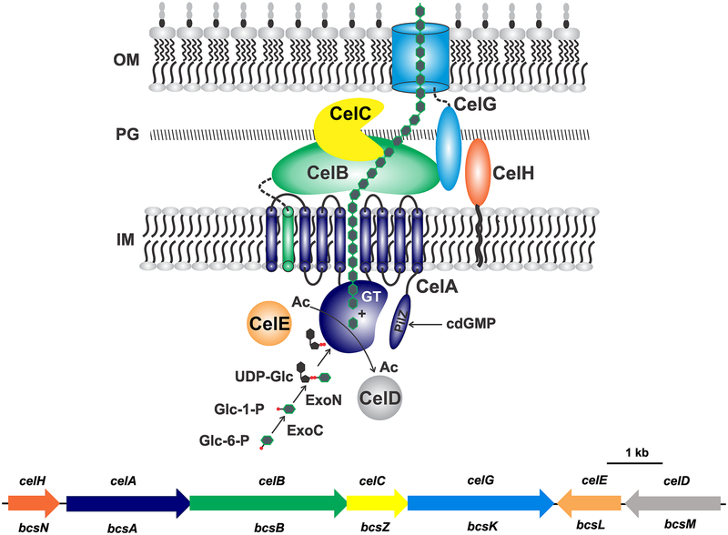
Figure 3. Cellulose: Genetic basis and biosynthesis model.
Predicted localization and mechanism of biosynthesis for cellulose in A. tumefaciens based on the generalized model (Römling and Galperin, 2015). Cellulose strand depicted as linked green hexagons. Cel protein names indicated in the figure. For CelA, GT is the predicted glycosyl transferase domain and PilZ is the cdGMP-binding domain. Black squiggle on CelH is a predicted lipid linkage. Gene colors match protein colors in the diagram; Cel names are above with corresponding Bcs nomenclature below. OM, outer membrane; PG, peptidoglycan; IM, inner membrane, Ac, acetyl groups; Glc-6-P, glucose-6-phosphate; Glc-1-P, glucose-1-phosphate; UDP-Glc, uridyl diphosphate glucose.

