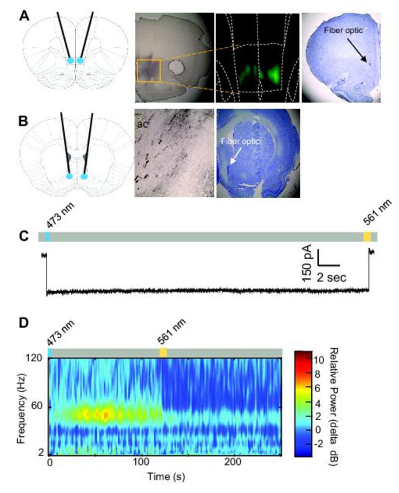
Figure 1. Confirmation of Stable Step-function Opsin expression and function.

A. Schematic of fiber-optic placement targeted at the IL (first panel). Anti-eYFP immunohistochemical staining from an SSFO-transduced rat (second panel). Fluorescent image shows SSFO expression in the IL (third panel). Cresyl violet staining shows a fiber optic terminating in the IL (fourth panel). B. Schematic of fiber-optic placement targeted at the NAshell (first panel). Anti-eYFP immunohistochemical staining of axonal fibers from the IL in the NAshell from an SSFO-transduced rat (second panel). Cresyl violet staining shows a fiber optic terminating in the NAshell (third panel). ac, anterior commissure C. Whole-cell voltage-clamp recording from an IL neuron transduced with SSFO. The representative trace shows inward current recorded following blue laser illumination (473 nm, 10 ms, blue rectangle). Trace shows return to baseline following yellow laser illumination (561 nm, 10 ms, yellow rectangle). D. In vivo electrophysiological recording from an IL neuron transduced with SSFO. Blue laser illumination (473 nm, 2 s, blue rectangle) enhanced 40–60 Hz activity, which was attenuated following yellow laser illumination (561 nm, 10 s, yellow rectangle).
