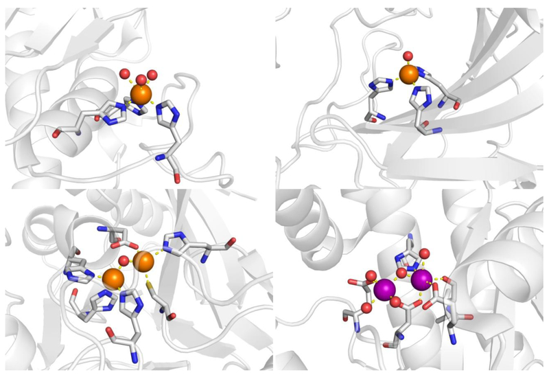
Figure 3.
Structures of metalloenzyme active sites used in this study. Metalloenzymes are shown as ribbons with coordinating residues shown in detail. Zn2+ is colored in orange spheres with Mn2+/Mg2+ in purple, and coordination bonds are displayed as yellow dashes. Upper left: MMP-12 (2OXU). Upper right: hCAII (1CA2). Bottom left: NDM-1 (3SPU). Bottom right: influenza endonuclease (5DES).

