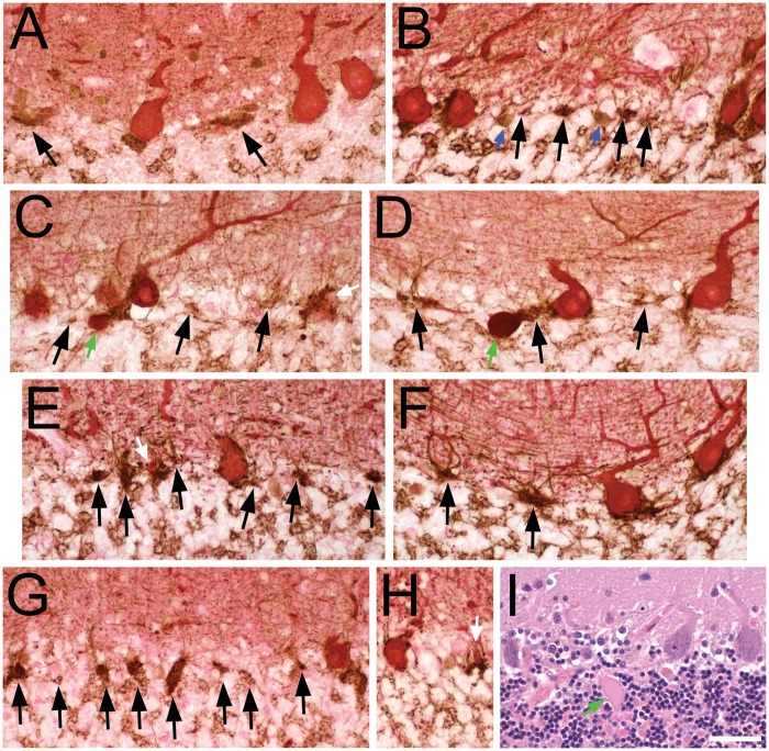
FIGURE 1.
Pathologic criteria for empty and full basket cells (A–H, calbindin D28k [CB] and glutamic acid decarboxylase (GAD) dual immunohistochemical staining), and histologic identification of Purkinje cells and torpedoes (I, Luxol fast blue-hematoxylin and eosin [LH&E]). (A–H) GAD-labeled pinceau (brown) have a characteristic overall triangular shape at the molecular layer-granule cell layer junction and at the base of PC soma (red CB-labeled). Perisomal basket cell processes extend vertically into the molecular layer, surrounding the PC soma (when present), and are of variable thickness and complexity. The presence of CB-stained PC cytoplasm classifies a basket as full, even if only a small portion of PC cytoplasm is identified (e.g. white arrows in C, E, H). Black arrows point to empty baskets, with a detectable portion of the pinceau, remaining basket processes in the adjacent molecular layer and lacking any detectable PC body. Granule cell layer interneurons (blue arrows in B) also stain with GAD and are distinguished by their rounder shape, presence of nucleus, and/or shorter branching processes. Axonal torpedoes (green arrows in C, D) stain red to reddish-brown with a dense glassy appearance, and are located beneath the brown GAD-stained pinceau, seen here in the upper granule cell layer. (A) Control. Three full baskets, 2 empty baskets; the perisomal basket processes are slender in this specimen. (B) Control. Four full baskets (one on right partially cut off), 4 empty baskets. (C) ET. Four full baskets, 3 empty baskets. (D) ET. Two full baskets, 3 empty baskets. (E) PD. Two full baskets, 6 empty baskets. (F) PD. Two full baskets, 2 empty baskets. (G) SCA2. One full basket, 8 empty baskets. (H) SCA1. Two full baskets. (I) LH&E-stained paraffin section from an ET case. Three PCs are counted; from left to right, the PC body has (i) only granular Nissl rich cytoplasm, (ii) a nucleus and a prominent nucleolus, and (iii) a nucleus lacking a nucleous. The axonal torpedo (green arrow) has a fusiform shape with glassy eosinophilic axoplasm. Scale bar (in I): 50 μm.

