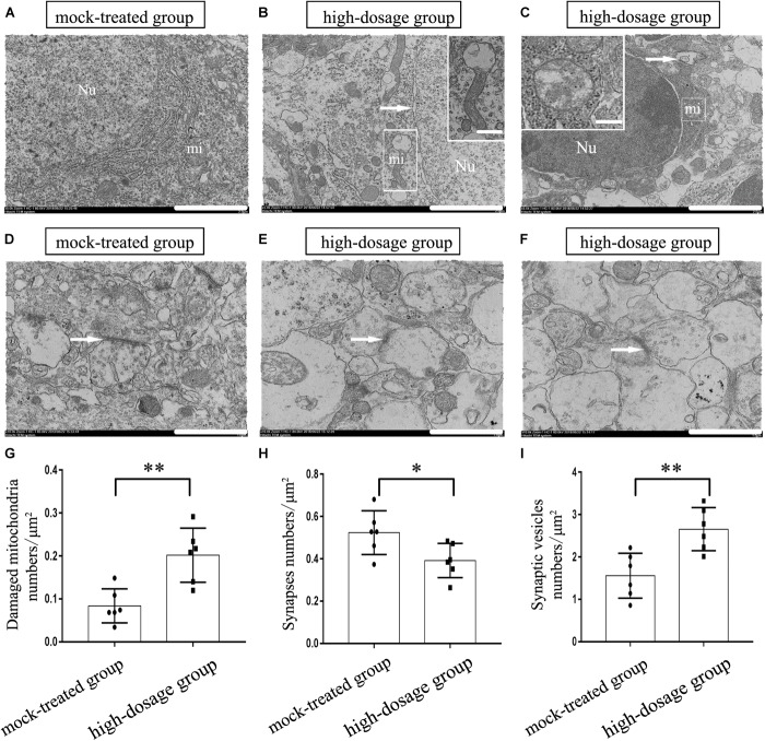
FIGURE 2.
Effects of gestational exposure to PM2.5 on the ultrastructure of neurons in 14-day-old mice offspring. (A–C) Neurons in mock-treated and high-dosage groups (Magnification is 5000×, bar = 2 μm, inset bar = 0.2 μm), the arrow shows the nucleus gap widening in (B) and autophagosome in (C), Neurons in high-dosage group displayed partial vagueness in mitochondrial cristae, vacuolar degeneration in mitochondrion. (D–F) Synapses in mock-treated and high-dosage groups (Magnification is 104×, bar = 1 μm). Nu, nucleus; mi, mitochondrion. (G) Numbers of damaged mitochondria statistical analyses per μm2 (t = 3.887, P = 0.003). (H) Numbers of synapses statistical analyses per μm2 (t = 2.455, P = 0.034). (I) Numbers of synapses vesicles statistical analyses per μm2 (t = 3.663, P = 0.003). ∗P < 0.05, ∗∗P < 0.01, compared with mock-treated group (n = 6).

