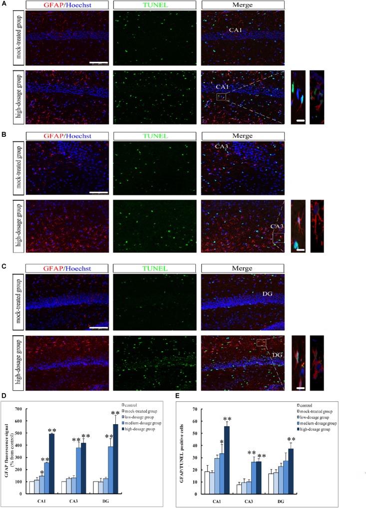
FIGURE 5.

Apoptosis of hippocampal astrocytes in 14-day-old mice offspring after exposure to PM2.5. (A–C) Laser confocal photographs of GFAP+/TUNEL+ in hippocampal CA1, CA3, and DG regions, respectively. GFAP staining (red), TUNEL staining (green) and nuclear staining (Hoechst, blue). Magnification is 400×, bar = 75 μm, inset bar = 10 μm. (D) GFAP fluorescence intensity as marker of reactive astrocytes (F = 511.345, P = 0.000, see CA1; F = 53.672, P = 0.000, see CA3; F = 58.273, P = 0.000, see DG). (E) GFAP+/TUNEL+cells (F = 30.690, P = 0.000, see CA1; F = 30.643, P = 0.000, see CA3; F = 10.686, P = 0.001, see DG). ∗P < 0.05, ∗∗P < 0.01, compared with mock-treated group (n = 6).
