Figure 2.
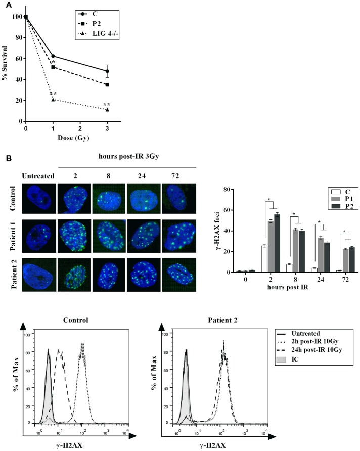
Cellular Response to DNA Damage. (A) Clonogenic survival assay in XLF-deficient fibroblasts. Cell survival following IR with γ-rays (1 and 3Gy) was assessed in primary fibroblasts from normal control (C) and patient (P2). LIG4-deficient fibroblasts (LIG4−/−) were used as a radiosensitive control. The results represent the mean and standard deviation of two separate experiments and are expressed as percentages of survival cells relative to unirradiated primary fibroblasts. (B) Top panel: Primary fibroblasts from control (C) and patients (P1 and P2) were irradiated with 3 Gy and fixed at given time points post-irradiation before staining with anti- γ -H2AX. Nuclei were stained with DAPI. Numbers of γ -H2AX foci per nucleus were determined at indicated time points after irradiation (average number of γ -H2AX foci per nucleus in 30 cells). Error bars represent the SD from 3 independent experiments. Bottom panel: γ-H2AX detection by flow cytometry was performed in PBMCs from P2 and control. Mean fluorescence intensities (MFI) are shown as histograms (unirradiated, 2 and 24 h) compared to isotype (IC). Persistence of γ H2AX signal at 24 h post-treatment in P2 is indicative of a general DNA repair defect. *p < 0.05, **p < 0.01.

