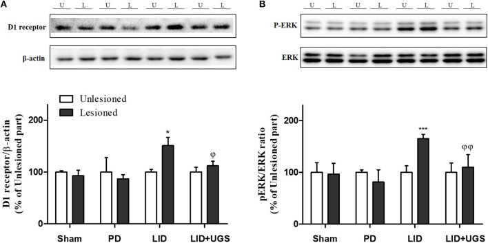
Figure 8.
UGS reduced the expression of D1R and the phosphorylation of ERK after development of LID. Representative band images and the quantification data of D1R (A) and p-ERK (B) for western blot analysis. Values of quantification data are given as the mean ± S.E.M. (N = 6). The significance of differences was analyzed by one-way ANOVA followed by Bonferroni's Multiple Comparison Test: *p < 0.05 and ***p < 0.001; mean values were significantly different from the unlesioned part of each group. ϕp < 0.05 and ϕϕp < 0.01; mean values were significantly different from the LID group.

