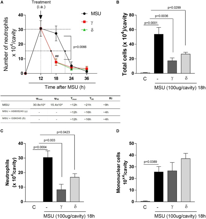
FIGURE 2.

Delayed treatment of PI3K-γ or PI3K-δ inhibitor in a MSU–induced gout inflammation induce timely resolution. Mice were injected with MSU crystals (100 μg) into the tibiofemoral joint and the treatment with intraarticular injection (i.a.,) of 50 μM of PI3K-γ or PI3K-δ inhibitor, 12 h after MSU injection. Cells were harvested from the articular cavity at 18, 24, and 36 h after MSU injection. The number of neutrophils and resolution indices were quantified (A). Of note: ψmax = maximal number of neutrophils, ψ50 = 50% of the maximum number of neutrophils, Tmax = 12 h, the time point when neutrophil numbers reach maximum; T50 MSU+AS605240 and MSU+GSK045 group ∼ 16 h, the time point when PMN numbers reduce to 50% of maximum; and resolution interval Ri MSU+AS605240 and MSU+GSK045 group ∼ 4 h, the time period when 50% PMN are lost from the articular cavity. Leukocytes counts 18 h after MSU injection (B) total leukocytes numbers, (C) neutrophils, (D) mononuclear cells. Results are expressed as the number of leukocytes per cavity and are shown as the mean ± SEM of five mice in each group from one experiment representative of two independent experiments. Significance was calculated using ANOVA followed by Holm-Sidak’s multiple comparison test. The exactly p-value are shown in the figure. ##means p value < 0.01 compared with 18 h MSU injected.
