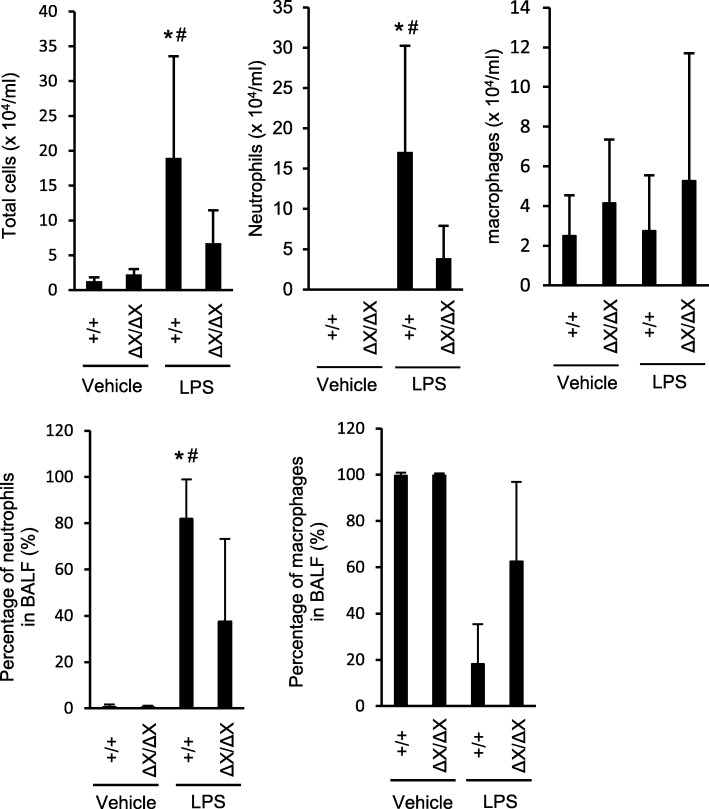
Fig. 2.
Effects of the PLCε genotypes on neutrophilic inflammation in LPS-induced ALI. BALF was collected in 24 h after i.t. administration of LPS or vehicle, cytospinned, and stained with a Romanowski stain (Diff-Quik). Leukocytes and neutrophils were counted under a microscope. n = 9 in the LPS-treated group, and n = 6 in the vehicle-treated group. *, p < 0.05 between control and LPS administration, #, p < 0.05 between PLCε genotypes

