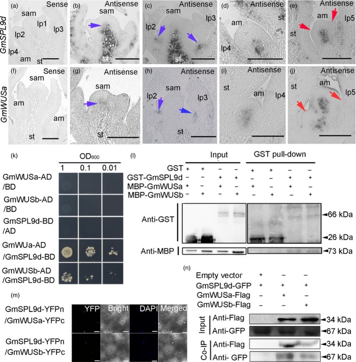
Figure 7.

GmSPL9d interacts with GmWUS. (a–e) In situ expression patterns of GmSPL9d in the shoot apical meristem (SAM) and axillary meristems (AMs). (a) Vegetative shoot apices at 15 days after emergence (DAE) were hybridized with a GmSPL9d sense strand control. (b) The GmSPL9d antisense probe signal appeared in the SAM (blue arrow). (c) The GmSPL9d signal appeared in the leaf axillary region (blue arrows) at the pre‐AM formation stage. (d) The GmSPL9d signal was detected in AM bumps. (e) GmSPL9d was expressed in established axillary buds. Red arrows show leaf primordia (LP). (f–j) In situ expression of GmWUSa in the SAM and at different stages of axillary bud formation in wild type. (f) The vegetative shoot apex of plants at 15 DAE was hybridized with a GmWUSa sense strand control. (g) The GmWUSa antisense signal appeared in the SAM (blue arrow). (h) A GmWUSa signal also appeared in the leaf axillary region (blue arrows) at the pre‐AM formation stage. (i) The GmWUSa signal detected in the AM bumps where GmSPL9d was expressed. (j) GmWUSa expression in an established axillary bud. Red arrows indicate LP. Note: lp1–5 represent LP at different developmental stages. st, stem. Scale bar, 50 μm. (k) Y2H assay validating the interaction of GmSPL9d with GmWUSa/b. (l) A GST pull‐down assay showing the interaction between GmSPL9d and GmWUSa/b. The sizes of GST tag, GST‐GmSPL9d fusion protein, MBP tag and MBP‐GmWUSa/b fusion proteins were indicated. (m) BiFC in tobacco leaves. Visible light indicates the interaction between GmSPL9d and GmWUSa/b in the nucleus. DAPI staining was used as a nuclear marker. Panels (left to right): YFP; bright; DAPI staining; merged channels. Scale bar, 25 μm. (n) Co‐IP of GmSPL9d‐GFP and GmWUSa/b‐flag. Transgenic plants expressing GFP were used as a negative control. The sizes of Flag tag, GmWUSa/b‐Flag fusion protein, GFP tag and GmSPL9d‐GFP fusion protein were indicated.
