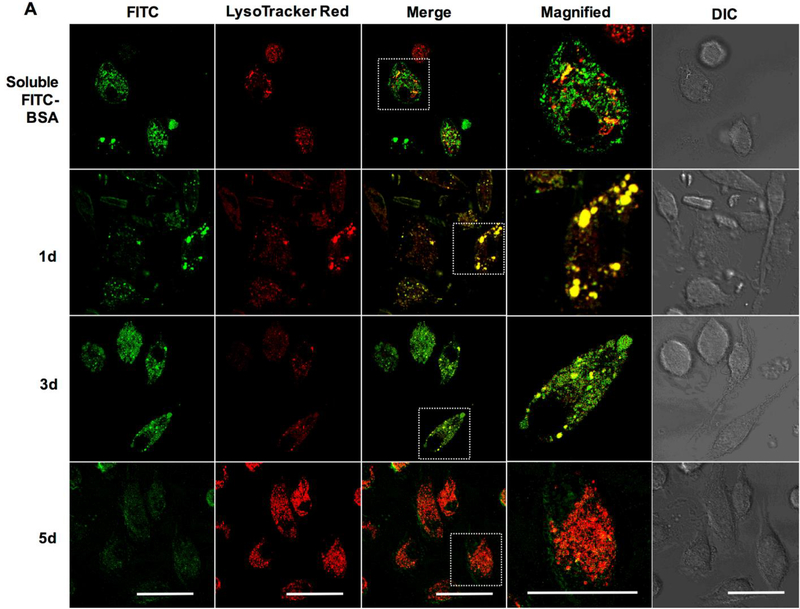
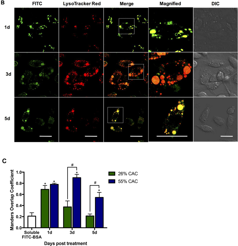
Figure 6. Time-dependent confocal microscopic analysis of fast- and slow-degrading FITC-BSA encapsulated Ace-DEX MPs co-localized to lysosomes.
Ace-DEX MPs composed of native Ace-DEX at (A) 26% cyclic acetal coverage (CAC) and (B) 55% CAC encapsulating FITC-BSA were delivered to bone marrow-derived macrophages at set time points, followed by live cell imaging using laser scanning confocal microscopy (40× oil immersion). Green: FITC-BSA, red: LysoTracker Red dye. Representative image of three independent experiments. Scale bar is 10 μm. (C) Using confocal images, co-localization was quantitatively measured by computing co-localization coefficients (Manders Overlap) using Image J and the plug-in JaCop for 26% (white bar) and 55% CAC (grey bar) Ace-DEX MPs, respectively. Values are reported as mean ± SEM. * indicates significance at p < 0.05 compared to non-treated control groups and # indicates significance between 26% and 55% CAC MPs.


