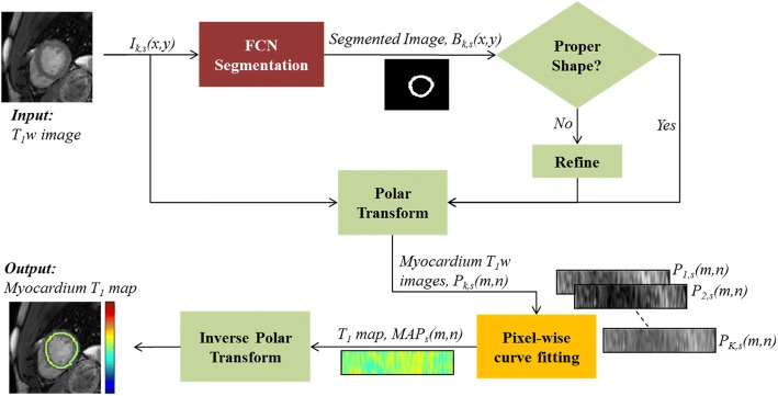
Fig. 1.
Pipeline for myocardium T1 map reconstruction. The myocardium in an input T1 weighted (T1w) image is first segmented using a fully convolutional neural network (FCN). The segmented myocardium is refined if needed (see text for details) and transformed into polar coordinates. All T1w images at a given slice are used to estimate the myocardium T1 map, which is displayed after applying inverse polar transformation

