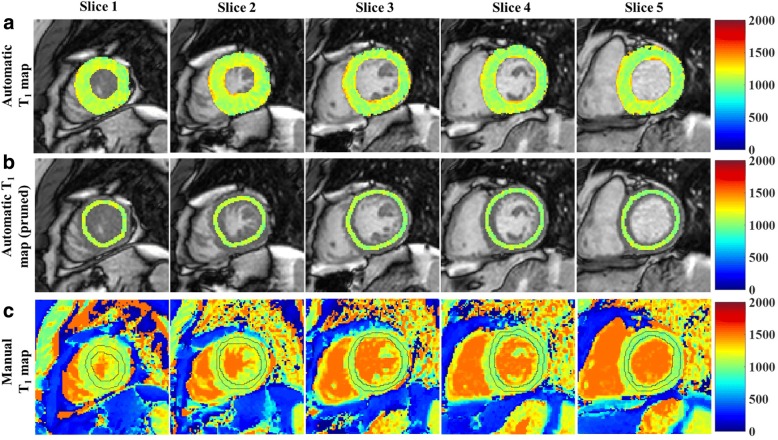
Fig. 5.
Myocardial T1 mapping at five short axial slices (apex to base from left to right respectively) of the left ventricle of one patient. Automatically reconstructed map before (a) and after (b) pruning overlaid on a T1 weighted image with shortest inversion time; (c) Manually reconstructed T1 map. The contours in (c) represent the myocardium region of interest manually selected by the reader

