Fig. 1.
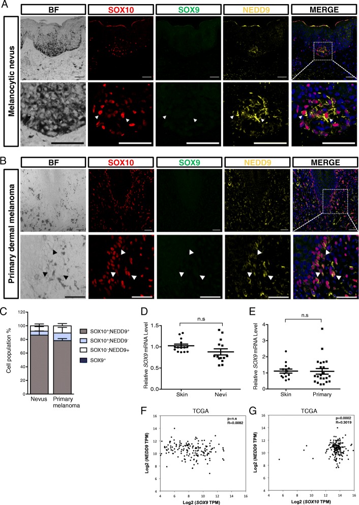
Co-expression of SOX10 and NEDD9 but not SOX9 in melanocytic nevi and primary dermal melanomas. a, b Representative images showing immunofluorescence for SOX10, SOX9, and NEDD9 in the skin sections of patients with benign melanocytic nevus (a) and primary dermal melanoma (b). White arrowheads indicate cells coexpressing SOX10 and NEDD9 but not SOX9. The dotted white box in the merged image indicates the magnified region with separate color channels shown in the lower panels. Cell nuclei were counterstained by DAPI (blue). Scale bars: 10 μm. c Quantification of the number of cells positive for the indicated markers in 12 melanocytic nevi and 14 primary dermal melanoma samples. d, e qPCR analysis of SOX9 expression in 14 healthy skin controls, 14 melanocytic nevi, and 22 primary melanoma samples. f Correlation expression analysis between SOX9 and NEDD9; SOX10 and NEDD9 (g) in melanoma patient samples obtained from Skin Cutaneous Melanoma dataset in TCGA (173 patients). Error bars represent mean ± SD. n.s, non-significant. The P-value and Pearson correlation coefficient are denoted on top

