Figure 3.

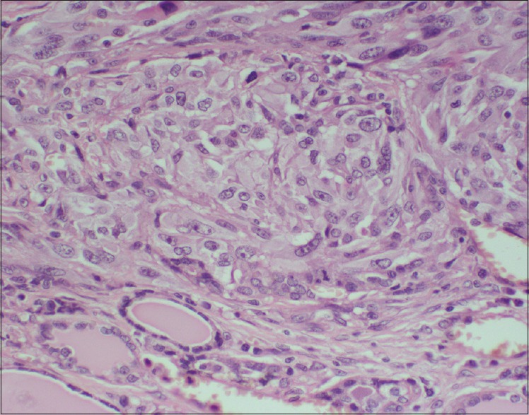
Microphotograph showing epithelial component with irregular vesicular nuclei and abundant cytoplasm (H and E stain, 400× magnification)

Microphotograph showing epithelial component with irregular vesicular nuclei and abundant cytoplasm (H and E stain, 400× magnification)