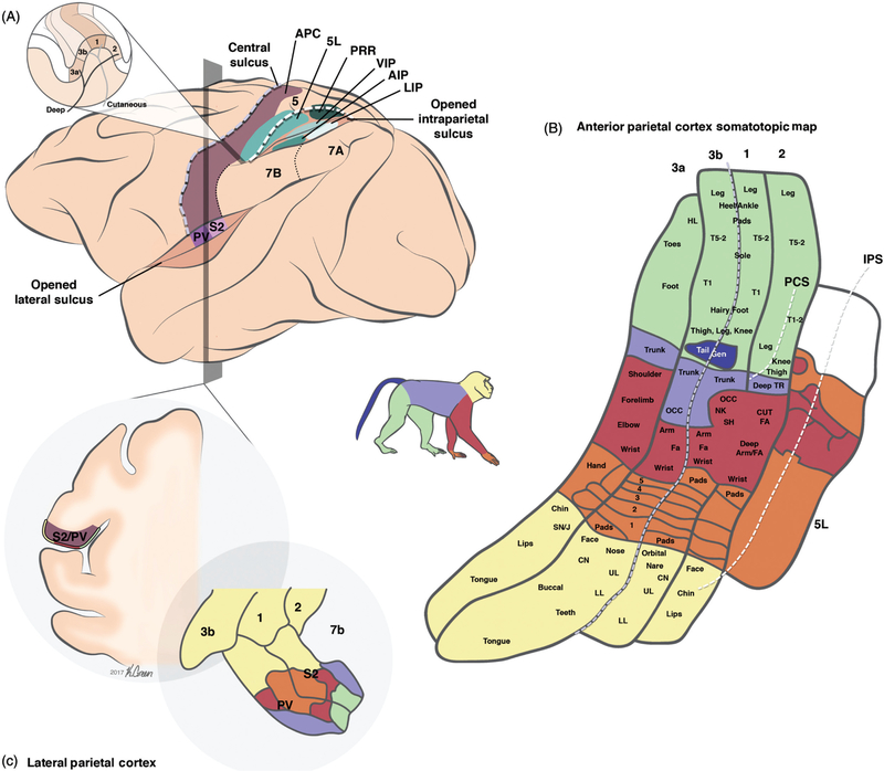
Figure 4.
Organization of somatosensory cortical areas. (A) A lateral view of the brain showing the different somatosensory areas in macaque monkey cortex. Adapted, with permission, from (198). Inset: Horizontal section of the postcentral gyrus at the level of the hand representation, showing the position of the different APC modules relative to the central and the intraparietal sulci. (B) Detailed view of the somatotopic representation of the body in the four fields of APC (areas 3a, 3b, 1, and 2) and in area 5L. Adapted, with permission, from (261, 332). (C) Coronal section showing the location of LPC in the lateral sulcus. Adapted, with permission, from (214). Abbreviations: Anterior parietal cortex (APC); second somatosensory area (S2); parietal ventral area (PV); parietal reaching region (PRR); anterior (AIP), ventral (VIP) and lateral (LIP) intraparietal areas; post central sulcus (PCS); intraparietal sulcus (IPS). Somatotopic map: Upper lip (UL); lower lip (LL); chin (CN); snout/jaw (SN/J); digits of the hand (1–5); (cutaneous) forearm ((CUT) FA); occiput (OCC); trunk (TR); toes (T1–5); hindlimb (HL).

