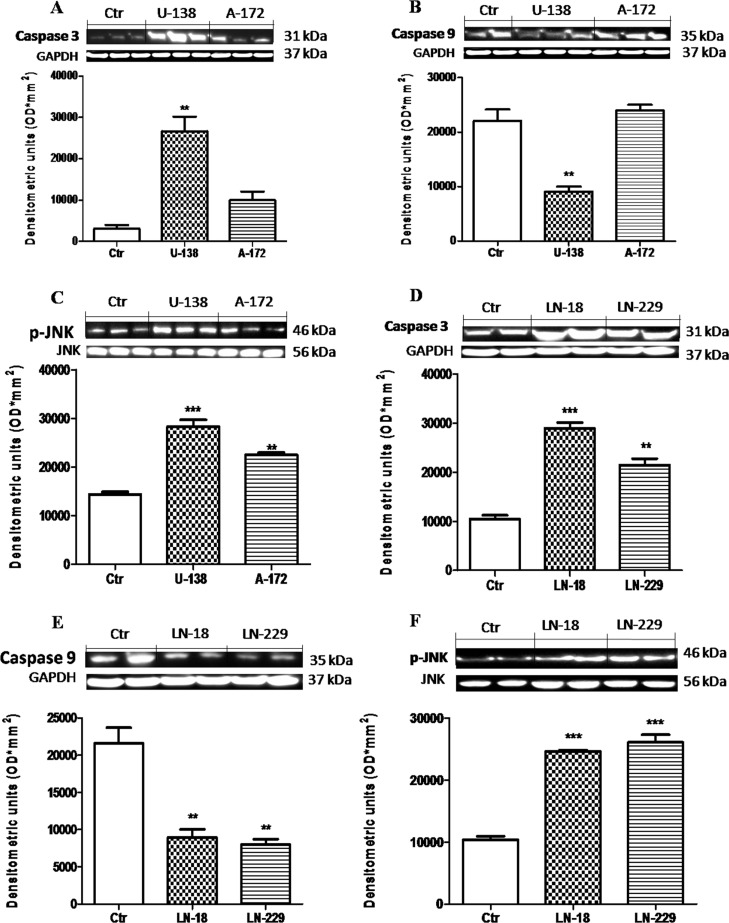
Figure 2. Apoptosic process in human glioblastoma cells.
A significant GBM cells apoptosis-resistance was observed in U-138MG, LN-18 and LN-229 cells, notably reduced in NHAcells (B and E). Conversely, Caspase-3 expression was significantly reduced in NHA cells, with a rising trend in A-172 and L-229 andsignificantly higher in U-138MG and LN-18 cells (A and D). Also, p-JNK expression was significantly higher in U-138MG, A-172, LN-18 and LN-229 cell lines, while lower p-JNK expression was highlighted in NHA astrocytes (C and F). Each data are expressed as mean ± SEM. One-Way ANOVA test followed by Bonferronipost test. *p < 0.05, **p < 0.01 and ***p < 0.001 vs Ctr. Caspase 3 (A) was detected in a stripped membrane. Therefore, the ubiquitary GAPDH band is the same for Dkk-3 and claudin-5 in Figure 1 (A and B respectively).

