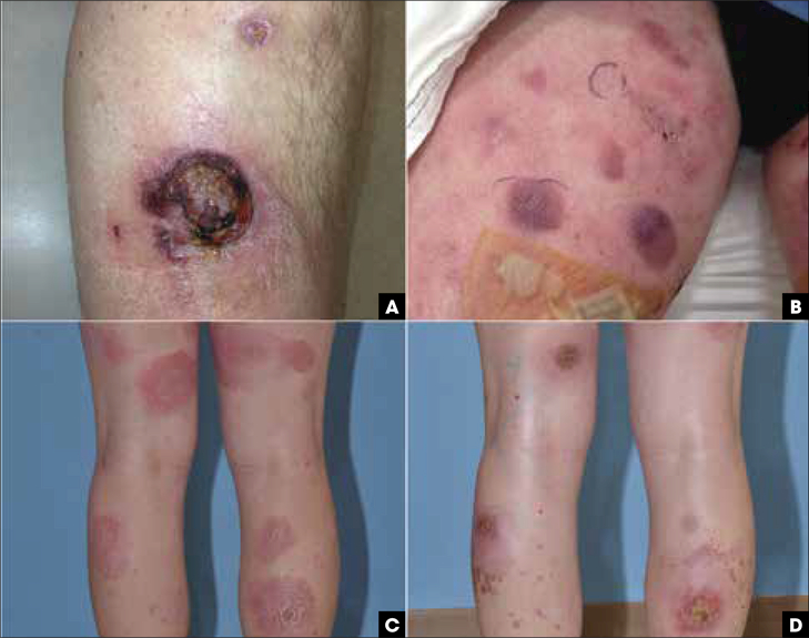
FIGURE 5.
Primary cutaneous γδ T-cell lymphoma, variable clinical presentation. (A) Ulcerated nodule on the anterior leg. (B) Violecous patches and plaques with focal erosions on the thighs. (C-D) Erythematous and scaly patches resembling mycosis fungoides on the legs that later evolved to ulcerated plaques.

