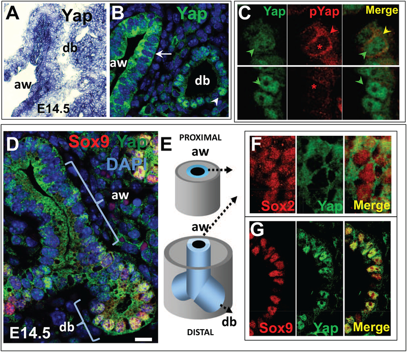
Figure 1. Differential activation of Yap in distinct populations of epithelial progenitors of the developing lung.
(A) In situ hybridization (ISH) of Yap showing widespread distribution of signals in airways (aw) and distal buds (db) of E14.5 mouse lung. (B) Immunofluorescence (IF) and confocal analysis of Yap (total) shows distinct nuclear (arrowhead, db) and cytoplasmic (arrow, aw) signals. DAPI (in blue) highlights nuclear staining. (C) Comparison of the pattern of Yap (total) and phosphorylated Yap (pYAP, S112-Yap phosphorylation) in E12.5 lung confirms pYap staining in the epithelium to be essentially cytoplasmic (red and yellow arrowheads); nuclear signals (green arrowheads in Yap staining) are absent in pYap-stained sections (asteriks). (D-G) Double IF analysis of Yap with markers of distal bud (Sox9) or airway (Sox2) cell fate during branching morphogenesis depicting overlapping Yap nuclear signals typically seen in Sox9-labeled distal buds largely absent in Sox2-labeled forming airways. The Yap cytoplasmic pattern is seen the vast majority of airway epithelial progenitors throughout the P-D axis of the lung (diagram in E; F-G). Scale bar in D, 12μm.

