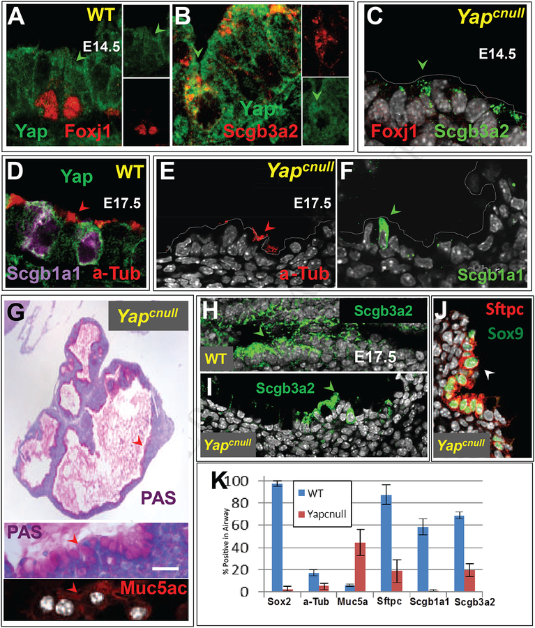
Figure 6. Yapcnull epithelium is unable to differentiate during lung development.
(A-B) Double IF of E14.5 WT lungs showing Yap pancytoplasmic signals (arrowheads) in ciliated (Foxj1) and secretory (Scgb3a2) cell precursors. (C) In E14.5 Yapcnull lungs no Foxj1 and only residual Scgb3a2 expression is present in the epithelium. (D) In E17.5 WT airways Yap-expressing cells undergoing differentiation express a-Tub and Scgb1a1 in ciliated and Clara cells, respectively (E-F). (H-I) By contrast E17.5 Yapcnull lungs show nearly no a-Tub, Scgb3a2 or Scgb1a1 staining and have abundant PAS and Muc5ac positive goblet-like cells in the epithelium. (J) Only rare clusters of double-labeled Sox9-Sftpc cells are present in E17.5 Yapcnull lungs. (K) Morphometric analysis showing the relative number of epithelial cells labeled with each differentiation marker in Yapcnull and WT lungs (mean ± SEM; n=3); Yap deficiency is associated with decreased expression of distal and proximal differentiation markers but increased Muc5ac expression. Scale bar, G, 40 μm.

