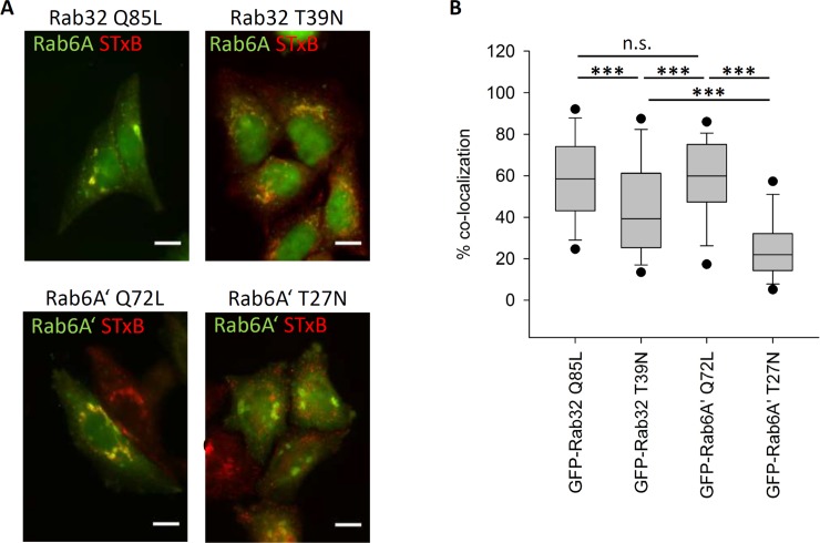
Fig 4. Rab32 influence on retromer mediated transport of Shiga toxin B.
(A) HeLa cells transfected with either GFP-Rab32 Q85L, GFP-Rab32 T39N (upper row), GFP-Rab6A’ Q72L or GFP-Rab6A’ T27N (lower row). 24 hours later cells were incubated with STxB-cy3 and pulse-chased for 60 minutes, followed by fixation in 4% PFA. Rab32-expressing cells were additionally stained by immunofluorescent labeling of Rab6A. (blue) to have a comparable Golgi marker to the GFP-Rab6A’ transfected cells. The Rab6A/A’ channels were depicted in green, STxB-cy3 in red. STxB co-localizing with the Golgi apparatus appears in yellow. GFP-Rab32 constructs are not visible. Scalebar = 10 μm (B) Quantification of STxB transport rates. Endogenous Rab6A or GFP-Rab6A’ was used to determine the localization of the Golgi apparatus. Transport rate is given as % co-localization of STxB with the Golgi apparatus. A lower % co-localization indicates a reduced rate of STxB transport. Rab32 Q85L: n = 127 cells, Rab32 T39N: n = 119 cells, Rab6A’ Q72L: n = 132 cells; Rab6A’ T27N: n = 131 cells.

