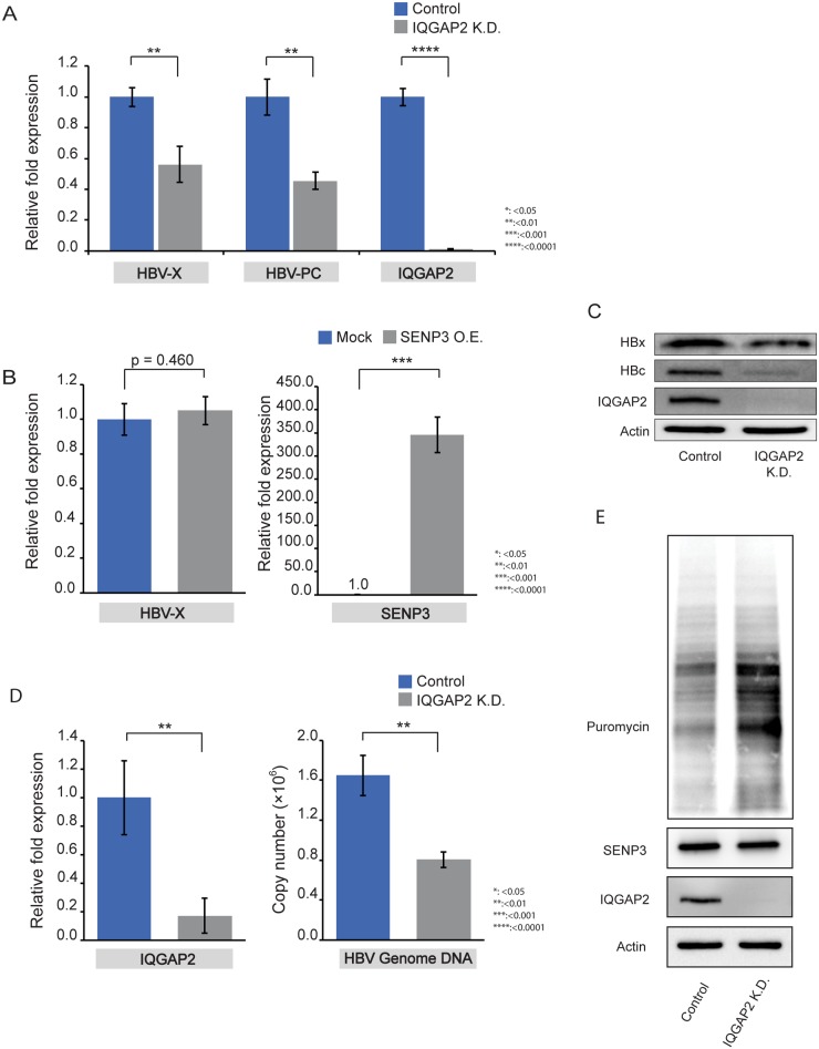
Fig 5. IQGAP2 silencing reduces viral gene expression and restores host translation.
(A) RT-qPCR measurements of HBV transcripts amplified by primers HBV-PC and HBV-X in HepG2.215 control cells and HepG2.215 IQGAP2 K.D. cells. Beta-actin was used as internal control; the data were expressed as mean±SD (n = 3). The statistical significance was assessed by Students’ unpaired t-test. (B) RT-qPCR measurement of HBV transcripts amplified by primer pair HBV-X in HepG2.215 IQGAP2 K.D. cells with transient transfection of pcDNA3 plasmid (Mock) and RGS-SENP3 plasmid (SENP3 O.E.). Beta-actin was used as internal control; the data were expressed as mean±SD (n = 3). The statistical significance was assessed by Students’ unpaired t-test. (C) Immunoblotting of HBx and HBc in HepG2.215 control and HepG2.215 IQGAP2 K.D. cells. (D) Left: RT-qPCR confirmation of IQGAP2 knockdown in HepG2-NTCP cells. Right: qPCR measurements of HBV genome copy numbers in supernatants of HepG2-NTCP cells (control and IQGAP2 K.D.) on Day 7 after HBV infection. Beta-actin (left) and HBV 1.3-mer WT replicon (Addgene Plasmid #65459) (right) were used as internal control. The data were mean±SD (n = 3) and the statistical significance was assessed by Students’ unpaired t-test. (E) Immunoblotting of puromycin-labelled proteins in HepG2.215-control and HepG2.215-IQGAP2 K.D. cells.

