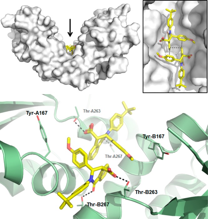
Figure 3.

X-ray structure of 1 in STING protein showing 2:1 binding and ligand interactions (PDB 6MX3). Top: STING is shown as a white surface, with 1 colored yellow. The inset shows a close-up view of the two copies of 1 from the direction indicated by the arrow. Bottom: close-up of polar interactions between 1 and side chains of Thr-263 and Thr-267 of both STING chains. Side chain atoms of Tyr-167 are also shown as sticks. Dashed lines indicate selected van der Waals interactions and hydrogen bonds.
