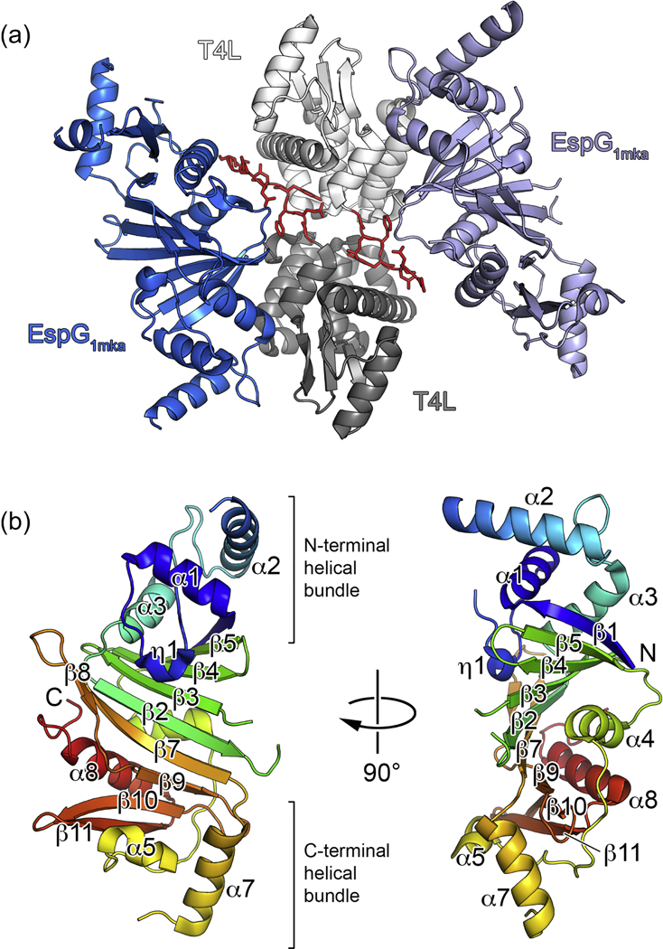
Figure 1. Crystal structure of M. kansasii EspG1 (EspG1mka).

(a) View of the two subunits of the T4L-EspG1mka fusion protein in the asymmetric unit. Chain A is shown in light grey (T4L) and light blue (EspG1mka), and chain B is shown in dark grey (T4L) and dark blue (EspG1mka). Residues corresponding to the TEV cleavage sequence are shown in red with side chains in stick representation. (b) A monomer of EspG1mka is shown in ribbon representation colored in rainbow colors from N-terminus (blue) to C-terminus (red). The N- terminal T4L fusion moiety is not shown for clarity.
