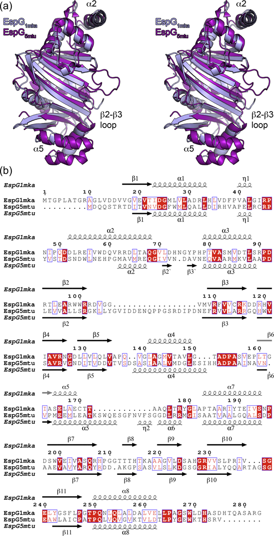
Figure 2. Structural comparison between EspG1mka and EspG5mtu.

(a) Stereo view of superposed EspG1mka and EspG5mtu crystal structures. The structure of the EspG5mtu monomer is derived from the trimeric EspG5mtu-PE25–PPE41–complex (PDB ID 4KXR, [26]). (b) Structure-based sequence alignment of EspG1mka and EspG5mtu. Secondary structure elements corresponding to the EspG1mka structure (PDB ID 5VBA) and EspG5mtu structure (PDB ID 4KXR) are displayed above and below the alignment.
