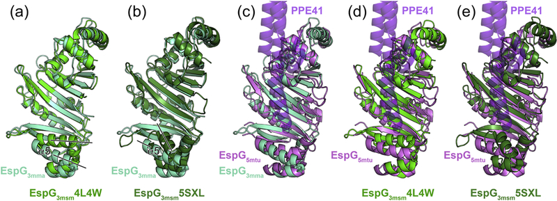
Figure 3. Crystal structures of EspG3 chaperones display variations in their putative PE-PPE binding region.

(a) Structural superposition of EspG3mma (aquamarine) and EspG3msm (PDB ID 4L4W, green). Black lines indicate differences in the orientation of the α5 helix. A stereo version is available as Supplementary Figure 4a. (b) Structural superposition of EspG3mma and EspG3msm (PDB ID 5SXL, dark green). A stereo version is available as Supplementary Figure 4b. (c,d,e) Structural superposition of EspG3mma, EspG3msm (PDB ID 4L4W), and EspG3msm (PDB ID 5SXL) with EspG5mtu (PDB ID 4W4I [27], violet) derived from the heterotrimeric EspG5mtu-PE25-PPE41 structure (PDB ID 4KXR [26]). PPE41 (purple) is shown in semi-transparent ribbon representation. PE25 is omitted for clarity as it is not in contact with EspG5mtu. Stereo versions of (c,d,e) are available as Supplementary Figure 5.
