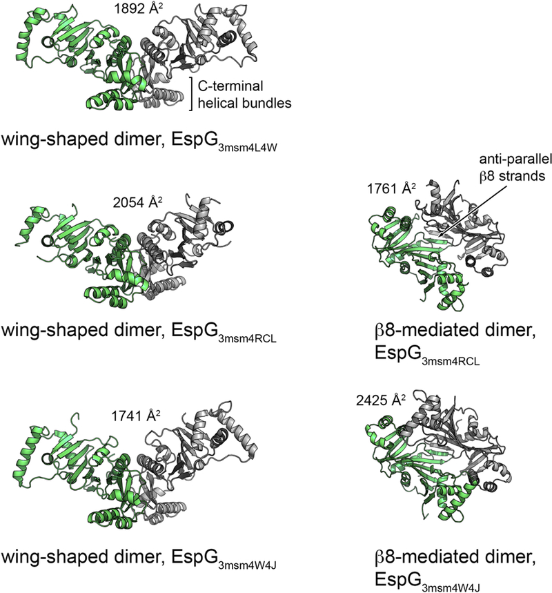
Figure 4. Cartoon representation of the common dimer structures observed in crystal forms of EspG3.

Superimposed subunits are in green with the buried surface area of the dimer interface indicated above the structure. The wing-shaped dimers are present in the asymmetric unit of EspG3msm (PDB ID 4L4W) and EspG3msm (PDB ID 4W4J) or generated by crystallographic symmetry in EspG3msm (PDB ID 4RCL). The β8-mediated dimer is present in the asymmetric unit in EspG3msm (PDB ID 4RCL) and generated by crystallographic symmetry in EspG3msm (PDB ID 4W4J [27]).
